The parasitoid wasp Rhyssa persuasoria (Linnaeus), also known as the giant ichneumon wasp, is used to illustrate the process of drilling wood with its ovipositor.
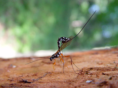
Photo 1. Rhyssa persuasoria Boris Hrasovec, Faculty of Forestry, Bugwood.org licensed under a Creative Commons Attribution-Noncommercial 3.0 License.
https://www.forestryimages.org/browse/detail.cfm?imgnum=9008010
Wasps are classified under the order Hymenoptera, suborder Apocrita. Apocrita is characterized by a narrowed waist known as a petiole, between the first and second abdominal segments. The first abdominal segment is also fused to the thorax. Hence the first abdominal segment, (after the petiole) we see in the field is actually the 2nd abdominal segment. Also, the larvae of all Apocrita lack legs, prolegs, or ocelli. The hindgut of the larvae also remains closed during development, with faeces being stored inside the body. Apocrita include wasps, bees and ants.
There are more than 150,000 species of wasps. Less than 1% of these species are eusocial. Paper wasps and hornets are examples of well-known social wasps. However, hornets can have upwards of 10,000 individuals in one nest. Thus, hornet encounters with man can sometimes lead to extremely painful or even fatal results. On the other hand, most of the wasps are solitary and seldom notice. Despite not being noticed, solitary wasps (because of their huge population numbers) are very important to Man as pollinators of plants and are one of nature’s most effective regulators of pest populations. Their larvae are parasitoid on a lot of Man’s agricultural insect pests.
Adults subsist on pollen and nectar and almost always hunt only to feed their larvae. With many species, individual wasps receive most of the nutrition they’ll ever get in life as larvae.
Creative Commons CC0 1.0 Universal Public Domain Dedication
As there are many parasitoid wasp species, there are also as many variations to the parasitoid lifestyles. We will look at Megarhyssa atrata to get an idea of what a parasitoid life can be like.
Megarhyssa atrata (M.atrata) is one of the Giant Ichneumon wasps belonging to the family Ichneumonidae, genus Megarhyssa (this genus has 32 species worldwide, 4 in North America).
M.atrata is a big wasp measuring 3.8 cm (from head to end of abdomen) for females. Males are smaller measuring about 3.5 cm. The body shape is like an extra-long cylinder. The females are mostly black in color except for a mostly yellow head, yellow feelers and legs. Part of the middle and hind legs are black. In contrast to males, the females have a long ovipositor. This is a hollow tube for egg laying, measuring 12cm to 15cm long (which is 3 to 4 times its body length). The males have more yellows on their bodies, black antennae, long segmented sausage-like abdomen and no ovipositor.
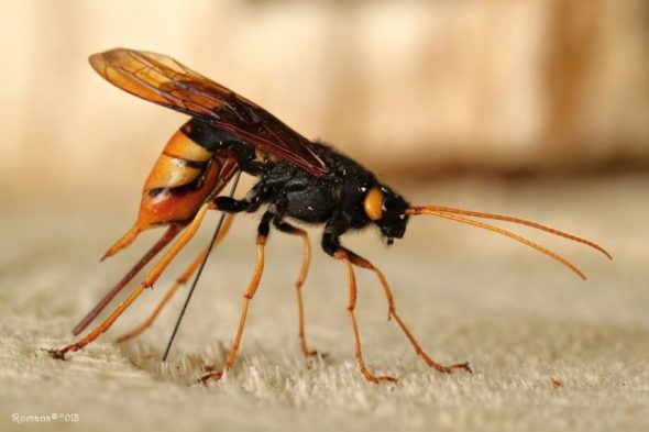
M.atrata is parasitoid on the pigeon horntail (Tremex columba) . Horntails, also known as sawflies, are wood wasps which lay their eggs in wood. Many horntail species are parasitised by parasitoid wasps. Photos of Urocercus gigas are used to illustrate horntail features.
CC-BY-NC-SA-3
Horntails or wood wasps are wasp-like flying insects but they do not have the narrowed waist of the true wasps. They are of the order Hymenoptera and family Siricidae (which includes 150 non-social species).
The name horntail is given because of the short, stout looking spike at the end of their abdomen. This horn is used to puncture the hard tree bark. Through this puncture, the female horntail then inserts her ovipositor (her second, and longer back end projection) and drills into the wood for about 2cm. If the wood is not too wet, she begins her ovipositing. She has hygroreceptors (microscopic sense organs called sensilla) at the tip of her ovipositor to help her. She usually deposits about 2 to 7 eggs, together with the white wood eating fungus, Cerrena unicolor, formerly known as Daedalea unicolor. This fungus is cultivated in a gland within her abdomen. This fungus predigest the wood and makes life easier for the horntail larvae when they hatch in 3-4 weeks. The horntail larvae grow within the dead or dying wood for 1 to 5 years. They then pupate near the bark. After about one month, they emerge as adults through round exit holes. Each adult horntail can lay up to 200 eggs. Horntails are found in south-eastern Canada, central and north-eastern parts of North America. M.atrata are specific parasitoids of pigeon horntails, thus they are found in the same area.
Between the months of June and September, M. atrata detects the location of a horntail larva deep within the wood of a dead or dying tree. It then drills in with its long ovipositor, maybe more than 10cm deep. It pierces the larva and injects paralysing venom. A single egg is then laid outside the horntail larva. The egg hatches and the wasp larva starts to eat up the horntail larva. The parasitoid larva pupates in spring and emerges in June to start the cycle.
Males (M. atrata) waiting for female to emerge through the bark. Video by Carl Barrentine Turtle River State Park North Dakota 14 Jun 2011
Life is not all rosy for the top “predator”. It may take more than an hour for the whole drilling process. During this whole time, they are stuck to the tree trunk. It is here that they are most vulnerable to attacks by big ants, lizards, chipmunks and birds. This explains why a lot of solitary ovipositors can be seen sticking in tree trunks.
In addition, after emerging from the pupa, not all adults are successful in digging a tunnel to the surface. Many fail and are entombed in the wood of the dead tree trunk.
The males usually emerge some days earlier. They are able to detect the locations of new emerging females, and males congregate around that spot. There are sometimes upwards of 10 males milling around the same spot. The competition is so stiff that some males are even seen placing their elongated abdomen down the hole that the female emerges from. When she emerges, males of other species will fly off. Only males of her own species will mate with her. She only mates once in her lifetime and lives for about 27 days after emergence. The males will fly off to look for another new emergent female to mate. They do not mate with older females.
The drilling process prior to ovipositing
M. atrata has first to locate the horntail larva, hidden deep within the tree trunk. It does this by detecting the sounds made by the larva as it chews its way through the wood. Sound waves are detected as vibrations through the solid medium of the wood. These vibrations are picked up by microscopic sensilla on the tips of the antennae, and larger, visible acoustic receptors (hairs and spines) on the legs of M. atrata. Modifications of wasp legs into hearing organs (large air spaces bordered by membranes) have been seen during dissections. Hence with this spatial audio detection system, M. atrata wasp can pinpoint the exact position of the horntail larva.
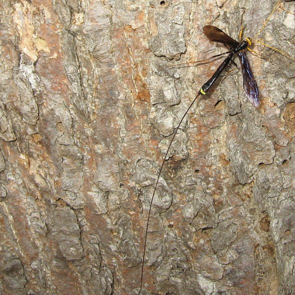
The next thing M.atrata wasp has to do is to set up the drill. This can be a complicated procedure. We use the wasp (Megarhyssa persuasoria) to illustrate this process.
Body length : 2 to 4 cm.
Ovipositor length : 2 to 4 cm (same length as its body)
https://www.youtube.com/watch?v=nxPST0qfvrg
Video by Miguel Angel Vicente Perez showing Rhyssa persuasoria ovipositing
Once the female has selected its target larva, Rhyssa persuasoria gets into position. It bends its body at the level of the petiole, so that its abdomen is now perpendicular to the surface of the tree trunk. At the same time, the end of the abdomen is curved downward, forming an “n” shape. The peak of the curve is between the 7th and 8th abdominal segment.
Each abdominal segment is covered by a skin, known as insect integument. This is mainly made of chitin, a polymer of glucosamine. In its pure form, it is a very tough and stretchable layer. For further protection, chitins in certain areas are hardened by a biochemical process called sclerotization. This is almost similar to the process of making melanin pigments in humans. In this process, very reactive quinones are produced, which causes an increase in cross-linkages between chitin polymers. At the same time, excess water is got rid of. This hardened chitin is used to protect the soft abdominal segment, dorsally called tergites and ventrally called sternites. Sclerotization is not an all or nothing phenomenon. It can take place to various degrees to produce varying degrees of hardness. The wasp abdominal sternites have less hardening and a soft median groove. This groove will receive the ovipositor when it is curved and pressed into it, during the ovipositing process. Along the length of the groove are tubercles which help to hold the ovipositor securely inside the groove.
The wasp ovipositor complex consists of the female T9 (9th abdominal tergum), two pairs of valvifers and three pairs of valvulae. Valvifers are hardened chitin plates, situated in the ventral region of the 8th and 9th abdominal segment. Together with the T9 tergum, these 5 chitin plates form a shoe-shaped structure which provides the point of attachment to the six sets of muscles that activate the ovipositor drill. All the valvulae are devoid of intrinsic musculature. Detailed anatomy is found here.
The 2nd valvifers (9th abdominal segment) extend caudally and give rise to the 3rd valvulae ( labelled as 3vv and form the ovipositor sheaths) and are anteroventrally articulated with the bulbous basal end of the 2nd valvulae (2vv). This is a loose type of ball and socket joint, which allows for erection of the ovipositor drill. This joint also allows for a small amount of rotational movement.
The 1st valvifers (8th abdominal segment) are continuous with the long and curved rami of the 1st valvulae (1vv).
The 1st and 2nd valvulae together form the actual tubular ovipositor. Each of the two pairs, consists of two tubular chitin structures. The two tubes of the 2nd valvulae are joined together by membranes and are fused into one tube at the tip. Hence, the 2nd valvulae function as a single unit. The 1st valvulae have similarly two tubular chitin structures and remain as individual functioning units, (the left 1st valve and the right 1st valve)
The interlocked 1st and 2nd valvulae enclose the egg canal and form the terebra (= ovipositor). The ventral surface of the 2nd valvulae is interlocked with both (the left and the right valves) of the 1st valvulae by two sublateral longitudinal tongues, which are T-shape in cross-section, called the rhachis. The left rhachis runs within a corresponding groove called the aulax (T shape in cross-section) along the dorsal surface of the left 1st valve. The right rhachis similarly runs in the aulax of the right 1st valve. This so-called olistheter system allows the three parts of the terebra to slide longitudinally relative to each other, while preventing their separation and thus maintains the integrity of the egg canal.
In the video by Miguel, the wasp erects its abdomen, bends the abdominal tip downwards and flexes its ovipositor upright to the drilling position. All the previous three changes occur simultaneously. This causes the separation of the proximal portion of the ovipositor from its protective sheaths. Note this separation in the video. All this maneuverings result in the ovipositor tip being applied to the surface of the tree trunk, in between the wasp’s two hind legs, giving increased stability. The wasp continues to press downward with its erected abdomen and bent tail. The proximal part of the ovipositor is now bent into a hook, like that of a walking stick. The rest of the ovipositor is also seen to be forcefully curved into the underside of the erected abdomen, pressed into the median abdominal groove and locked into place. This posture results in the long ovipositor being held securely, thus facilitating the adjustment of her drilling angle. This posture is also stabilized by the extra big and long hind legs, planted each on either side of the drill. In the initial posturing, if the ovipositor is too long to get into proper position, some wasps bring their hind legs closer together. This will elevate the height of the “abdominal crane” and enable the ovipositor to be placed upright. However, this has the disadvantage of reducing the stability of the whole drill setup and the wasp may, and does topples over. If that happens, it will have to start all over again.
Start of the drilling process. This actual drilling is not seen in the video. We can only see the result itself, when the long and flexible, hair-like, thin, ovipositor is being pushed 3 to 4 cm deep into the dry, hard wood.
How does the drilling process work? Scientists have spent many hours using dissection, light microscopy and scanning electron microscopy to tease out an answer(s). Some of their research papers are referenced below.
During the drilling process, the right and left valves (of 1st valvula) are deployed in rapid alternating downward piercing and upward rippling motions. There are saw teeth, about 5 in number, on the ventral aspect of the valves, decreasing in size towards the tip.
Left 1st valvula: On the downward stroke, it pierces the cellulose cell wall of the tree wood. On the upward stroke, it rips open the wood cell, and at the same time the more proximal and larger upward pointing, scoop-like saw tooth, will gouge out the wood on the side, enlarging the tunnel.
Once the cell wall is ripped open, the 2nd Valvulae are pushed into the wood cell by the gentle constant pressure exerted by the whole abdominal and leg musculature, with the claws at the end of the legs providing stabilising anchorage. The tips of the 2nd valvulae now rest on the roof of the next layer of wood cells. Too much downward pressure may result in the ovipositor buckling and breaking. However, buckling cannot always be avoided, as when an area of hard wood is met. Thus, some parts of the long tubular ovipositor cuticle have a rich addition of Resilin, so that buckling will not result in breakage. This is one of the most elastic proteins known.
Right 1st valvula: On the up-stroke of the left 1st valvula, the right valvula moves down and pierces the roof of the next layer of wood cells. It then moves upward to rip open the roof of this 2nd layer of wood cells. This upward motion will also allow the bigger serrations situated higher up the valvula to gouge the wood along the sides of the tunnel. While doing this, it provides a counter force to aid the downward stroke of the left 1st valvula. This action reduces the overall forces needed to push the ovipositor downwards.
The cyclic motion, whereby one valve is pulled backwards and anchored, whilst the other is pushed further into the wood, enables the forward motion to be achieved with a near-zero net force, minimising the chance of the long ovipositor buckling.
In the next article “Parasitoid wasp 3”, another wasp (Megarhyssa macrurus), with ovipositor that is 3-4 times the length of its body, will be described. This wasp takes additional steps to deal with problems associated with an extremely long ovipositor.
Body length : up to 5 cm
Ovipositor length : up to 13 cm
Article by Michael Wong
Photo credits :-
Photo 3. Author: Romana Plačková [Other photographs by this author]
Determination author: Romana Plačková [Determination history and verification]
Origin of image Slovakia, Malá Fatra 5 July 2013
Author rights ?
| CC-BY-NC-SA Creative Commons Attribution NonCommercial ShareAlike License 3.0 [Information]
|
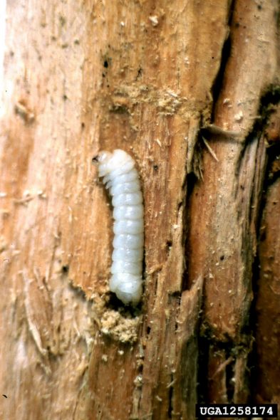
Photo 4. giant wood wasp (Urocerus gigas) larva
giant wood wasp
Insecta (Hexapoda) > Hymenoptera > Siricidae
Urocerus gigas L.
Photographer: Stanislaw Kinelski,
Contact: Lidia Sukovata, Polish Forest Research Institute
Descriptor: Larva(e)
Image Citation: Stanislaw Kinelski, , www.forestryimages.org
Image Use: This image may be copied and used, in whole or in part, for any non-profit, educational purpose provided that all reproductions bear an appropriate credit. Any commercial or other use of the image requires the written permission of the photographer or contact organization, and Forestry Images.
References:
1) Ovipositor of the braconid wasp Habrobracon hebetor: structural and functional aspects. June 2021,
- Journal of Hymenoptera Research 83:73–99
Authors: Michael Csader, Karin Mayer, Prof. Dr Oliver Betz, Stefan Fischer, Benjamin Eggs
2) Mechanisms of ovipositor insertion and steering of a parasitic wasp (Research article)
PNAS : Proceedings of the National Academy of Science of the United States of America.
PNAS September 12, 2017 114 (37) E7822-E7831; first published August 28, 2017; https://doi.org/10.1073/pnas.1706162114
View ORCID ProfileUroš Cerkvenik, Bram van de Straat, Sander W. S. Gussekloo, and Johan L. van Leeuwen
Experimental Zoology Group, Wageningen University and Research, 6708WD Wageningen, The Netherland
3) Structure and function of the musculoskeletal ovipositor system of an ichneumonid wasp (research article)
4) Wikipedia – hymenoptera, Wasp, parasitoid wasp, chitin, resilin, Rhyssa persuasoria, horntail,
5) www.earthlife.net/insect/anatomy