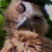
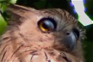
In early April 2010, KC Tsang made a video of a sleepy juvenile Buffy Fish Owl (Ketupa ketupu) roosting on its perch (left). It was around 1530 hours and the owl was preening its upper feathers. During the short period it was preening, the eyes showed a combination of opening, closing and movements of the nictitating membrane.
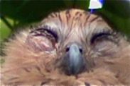
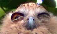
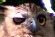
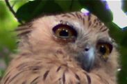
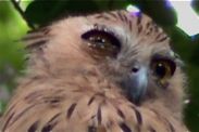
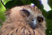
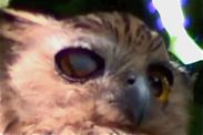
The images below best illustrate this: left shows both eyes closed, centre shows one eye about to close while the other already closed and right shows one eye fully open while the other has the upper eyelid blinking and the nictitating membrane over it.
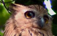
The video provides an opportunity to study the opening and closing of the eyes. As typical of birds, closing of the eyes involves the lower eyelids. They move upwards to cover the eyes. The images below clearly illustrate the stages of closing of the right eye. All the time the left eye remains open.
Similarly, the video clearly shows the appearance of the nictitating membrane as the upper eyelid is flicked downwards. The nictitating membrane appears from the inner upper corner of the eye to move diagonally towards the lower outer corner. The images below illustrate the stages in the right eye.
The bird’s eye has an upper and a lower eyelid as well as a nictitating membrane or third eyelid. According to Evans & Heiser (2004), this membrane “moves sideways across the eye, at right angles to the regular eyelids, cleaning the eye’s surface and keeping it moist.” In an earlier post we showed the nictitating membrane moving sideways in the case of the Plaintive Cuckoo (Cacomantis merulinus). However, in Buffer Fish Owl, the membrane moves diagonally, as also shown in an image posted earlier. Long (1998) has confirmed this, describing the movement “from the inside corner diagonally to the outside corner…” Furthermore, Long’s (1998) take on the eyelids… “The upper eyelids are used in blinking movements, which helps clean the eyes. The lower eyelids are pulled up over the eyes during sleep.”
Images are video grabs copyright of KC Tsang. The video can be viewed HERE.
References:
1. Evans, H. E. & J. B. Heiser, 2004. What’s inside: Anatomy and physiology. In: Podulka, S., R. W. Rohrbaugh Jr & R. Bonney (eds.), Handbook of bird biology. The Cornell Lab of Ornithology, Ithaca, NY. Pp. 4.1-4.162.
2. Long, K., 1998. Owls: A wildlife handbook. Johnson Books, Boulder. 181 pp.




3 Responses